


Understanding DNA Repair: Visualizing Tumour Suppressors in Action.
The genome is constantly threatened by damaging agents, and accumulated damage can lead to cancer and other genetic disorders. Cells have evolved an intricate…
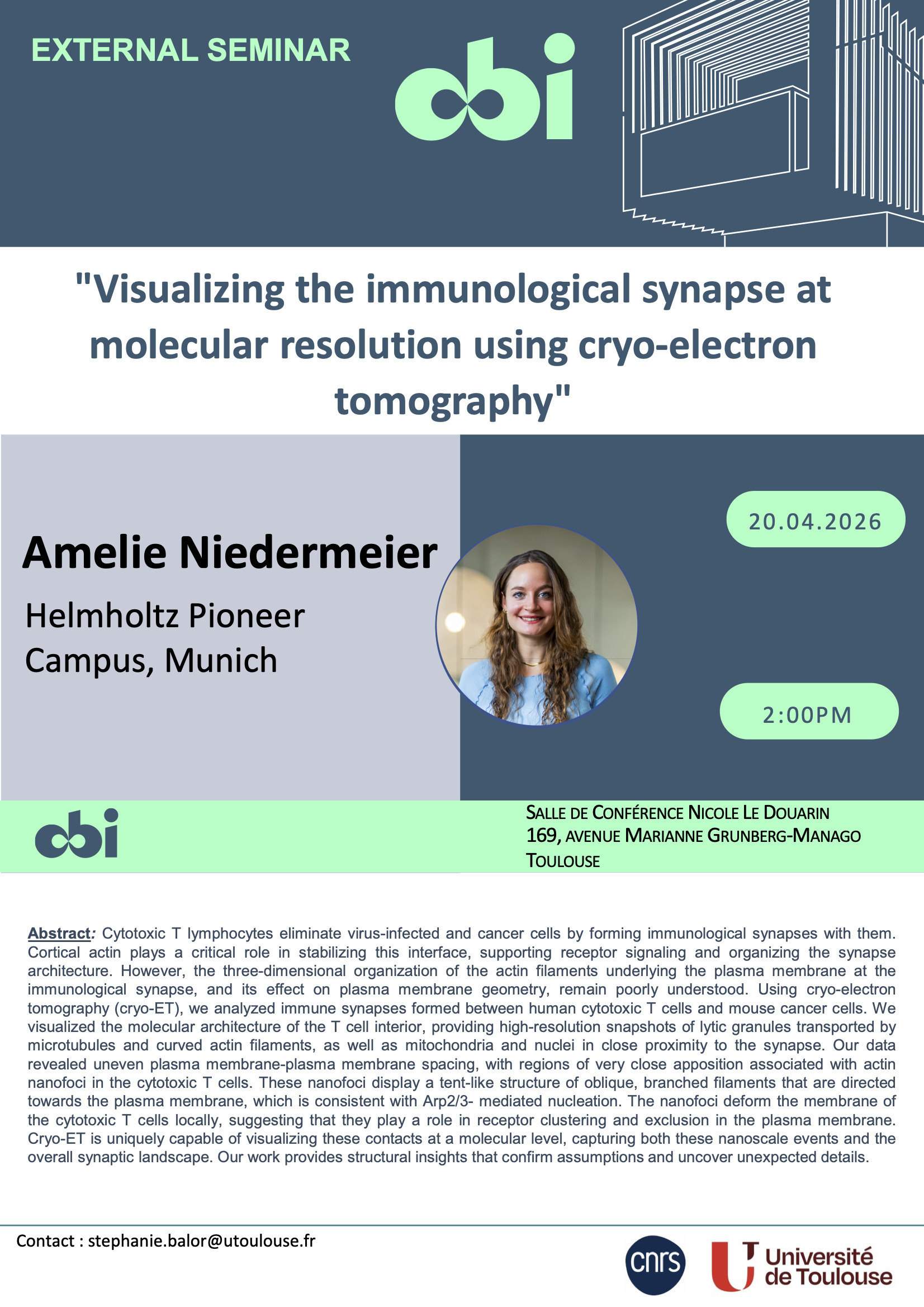
Cytotoxic T lymphocytes eliminate virus-infected and cancer cells by forming immunological synapses with them. Cortical actin plays a critical role in stabilizing this interface, supporting receptor signaling and organizing the synapse architecture. However, the three-dimensional organization of the actin filaments underlying the plasma membrane at the immunological synapse, and its effect on plasma membrane geometry, remain poorly understood. Using cryo-electron tomography (cryo-ET), we analyzed immune synapses formed between human cytotoxic T cells and mouse cancer cells. We visualized the molecular architecture of the T cell interior, providing high-resolution snapshots of lytic granules transported by microtubules and curved actin filaments, as well as mitochondria and nuclei in close proximity to the synapse. Our data revealed uneven plasma membrane-plasma membrane spacing, with regions of very close apposition associated with actin nanofoci in the cytotoxic T cells. These nanofoci display a tent-like structure of oblique, branched filaments that are directed towards the plasma membrane, which is consistent with Arp2/3- mediated nucleation. The nanofoci deform the membrane of the cytotoxic T cells locally, suggesting that they play a role in receptor clustering and exclusion in the plasma membrane. Cryo-ET is uniquely capable of visualizing these contacts at a molecular level, capturing both these nanoscale events and the overall synaptic landscape. Our work provides structural insights that confirm assumptions and uncover unexpected details.
The genome is constantly threatened by damaging agents, and accumulated damage can lead to cancer and other genetic disorders. Cells have evolved an intricate…

The ~ 100 imprinted genes in mammalian genomes are mono-allelically expressed based on their parental origin (i.e. they are only active on the chromosome…

Mouse Cortex development

La Communauté Municipale de Santé est heureuse de vous présenter cette exposition/vente. Huit artistes toulousains; David Kassar, Député, Pitu, Vincent…

Événement réservé aux demandeurs d'emploi inscrits à l'agence Toulouse Cartoucherie ! Vous avez 50 ans et plus. Faites le point sur votre situation, votre…

Adieussiatz brave monde! Retrouvons-nous le lundi 20 avril à partir de 20h pour chanter en polyphonies, en occitan, en français, en basque... Au plaisir de s'y…

Vous recherchez un emploi dans le secteur du Bâtiment ? Venez rencontrer AIDAL 31 (ETTI- SIAE) agence de travail temporaire d'insertion professionnelle dans…

Capemploi Haute Garonne vous propose un atelier d'informations avec un agent de Tisséo, qui vous présentera les différentes offres tarifaires spécifiques aux…

Les métiers des Services à la Personne recrutent ! Vous recherchez un emploi ? Vous débutez dans le secteur ou souhaitez vous reconvertir ? Venez rencontrer…

Le Sangha Hotel à la Cartoucherie recrfute des femme/homme de Chambre débutants pour la saison (CDD 4 mois), Mais aussi des Serveur/se et Chef(fe) de rang pour…

Découvrez les collections insolites du lieu à travers un parcours dynamique, prétexte à se questionner en famille sur les missions des musées en général !…
Commentaires
Aucun commentaireUne question sur les horaires, l'accès ou le programme ? Envie de partager vos impressions ? Échangez avec la communauté By Night.